Scientists at the National Institutes of Health (NIH) analyzed postmortem samples taken between April 2020 and March 2021. They took extensive samples of the nervous system, the brain, in 11 of the patients.
RNA and virus viable in organs
All of the patients have died with COVID-19 and none have been vaccinated. Blood plasma from 38 patients tested positive for SARS-CoV-2, 3 tested negative and plasma not available to the other 3.
30% of patients were women and the median age was 62. 5 years. Twenty-seven patients (61. 4%) had 3 or more comorbidities. The median period between onset of symptoms and death was 18. 5 days.
The research showed that SARS-CoV-2, as expected, basically inflamed and ruptured the respiratory tract and lung tissue. But the researchers also discovered viral RNA in 84 different places and structural fluids, and in one case, isolated viral RNA 230 days after the patient’s symptoms began.
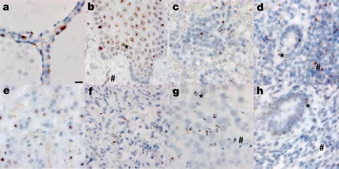
The researchers detected SARS-CoV-2 RNA and proteins in the hypothalamus and cerebellum of one patient and in the spinal cord and base ganglia of two other patients. But they found little damage to brain tissue, “despite a higher viral load. “
The researchers also isolated the viable SARS-CoV-2 virus from the internal and external tissues of the airways, adding the brain, heart, lymph nodes, gastrointestinal tract, adrenal gland, and eyes. They removed the virus from 25 of the 55 samples tested. (45%).
The authors wrote, “We demonstrated replication of the virus at several non-respiratory sites in the first two weeks after symptom onset. “
They add: “Our short autopsy intervals, a comprehensive standardized technique for tissue collection, brain dissection before fixation, preservation of tissue into RNA later, and instant freezing of new tissue allowed us to stumble and quantify SARS-CoV-2 RNA levels. with maximum sensitivity by [polymerase chain reaction] and [in situ hybridization], as well as to isolate the virus in mobile culture of several non-respiratory tissues, adding the brain, which are notable differences with other studies.
Possible ramifications of COVID
The study’s lead author, Daniel Chertow, MD, MPH, said in an NIH news release that prior to the work, “the general thinking was that SARS-CoV-2 was primarily a respiratory virus. “
The discovery of viral presence in the frame, and sharing those findings with colleagues a year ago, helped scientists explore a relationship between highly inflamed frame tissues and “prolonged COVID,” or symptoms that persist for weeks and months after infection.
Part of a Paxlovid RECOVER trial expected to begin in 2023 comes with an extension of the post-mortem paintings highlighted in the Nature study, according to co-author Stephen Hewitt, M. D. , Ph. D. , who sits on the RECOVER Project Steering Committee. Autopsies from the RECOVER trial come with other people who were vaccinated and inflamed with fear variants. knowledge that was not had in yesterday’s study.
Please indicate the appropriate maximum category to facilitate the processing of your request
Thank you for taking the time to provide feedback to editors.
Your opinion is for us. However, we do not guarantee individual responses due to the large volume of messages.